


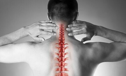
Cervical osteochondrosis, or spondylosis, results from changes in the shape and structure of the vertebrae.Although the cervical region is quite short compared to the total length of the spine, it is perhaps the most important part of the spine.Each pair of adjacent vertebrae forms intervertebral foramina through which nerve roots emerge and are directed to every muscle and organ in the upper half of the body.Vital vessels that supply blood to the brain pass through other openings - in the lateral processes of these vertebrae.
Causes of osteochondrosis of the cervical spine
The causes of osteochondrosis are:
- injuries,
- “sedentary” work on a monitor located below eye level,
- physical work associated with carrying heavy loads,
- drive a car for a long time,
- work “on the phone” without using remote devices (in this case, the operator presses the receiver to his ear with his shoulder)
- constitutional characteristics (torticollis, congenital changes in the cervical vertebrae, short neck)
Formation of pathological changes in the vertebrae
With osteochondrosis, small sharp points begin to form on the edges of the vertebral bodies, which can injure nearby structures.Most often, this occurs in response to excessive load on the cervical spine, and is not only the result of "aging" of the intervertebral joints (recall that osteochondrosis was previously considered a degenerative and natural "age-related" disease, like osteoarthritis).As the disease progresses, the endplates become denser and the height of the intervertebral discs decreases.These discs normally act as shock absorbers between the vertebrae, and prevent, among other things, damage to the spinal roots.With progressive osteochondrosis, a protrusion (herniation) of the nucleus pulposus of the intervertebral disc occurs, on which, during the disease, increasing pressure is exerted while the ligaments that “hold back” from all sides are weakened.This hernia can also compress spinal structures and cause neurological manifestations of the disease.
What are the symptoms of cervical osteochondrosis?
Osteochondrosis of the cervical spine with pain syndrome
Any pain in the neck raises suspicion of cervical spine pathology.Depending on the increasing intensity of the pain syndrome, they are divided into 4 stages, at the first the patient feels numbness, tingling, a feeling of "tightness" in a certain muscle group, at the fourth stage - the most severe - the pain is so intense that it leads to immobility of the patient and loss of performance.
In addition to pain in the cervical and occipital region, the patient notes "referred" (radiating) pain in the upper extremities and lateral subscapular areas of the chest.
Osteochondrosis of the cervical spine with radicular syndrome
The involvement of nerve roots in the process is indicated when pain, numbness and tingling spread to the lower jaw, upper back, forearm and fingers.At the same time, the patient draws attention to the fact that he “seemed to rest” his hand and slept uncomfortably.There is morning stiffness in the finger joints, which lasts no more than 10-15 minutes.With the development of radicular syndromes, upon examination, a decrease in muscle strength of the upper extremities may be noted.
Osteochondrosis of the cervical spine with “vertebral artery syndrome”
The involvement of blood vessels in the process (compression by a hernial protrusion or osteophyte) is indicated when the patient complains of frequent attacks of headaches, especially after being in a certain position for a long time, when throwing the head back (for example, when swimming breaststroke), in case of tinnitus and dizziness.This clinical situation is clearly identified using ultrasound (with “Doppler mapping mode”).Ultrasound reveals tortuosity of the vertebral arteries and narrowing of their lumen.In this case, we can talk about surgery, since a pronounced change in blood flow in the vertebral arteries is a risk factor for stroke.
Osteochondrosis of the cervical spine with “cardiac syndrome”
This syndrome forces the patient to first turn to a cardiologist, since the main complaints are pain in the left half of the chest, the subscapular region, which weakens or intensifies when performing physical activity or changing body position.After excluding myocardial infarction and other heart diseases, the patient is admitted under the supervision and treatment of a neurologist and orthopedist.
Diagnosis
To clarify the diagnosis, four methods are used: x-ray, ultrasound, computed tomography and magnetic resonance imaging.
The most accessible method remains cervical spine radiography;the most informative is the lateral projection radiograph (“lateral view”).This method allows, as a first approximation, to determine the presence of injuries and significant structural changes in the vertebrae.
An ultrasound examination (ultrasound) is carried out to clarify the condition of the vertebral arteries.This method makes it possible to determine whether blood flow is impaired and, if so, to what extent and what types of obstacles have appeared and where they are located.
Computed tomography (CT).Allows you to more accurately assess the condition of bone structures, the degree of density of bone tissue and allows you to see osteophytes (bone growths) smaller than with x-ray.
Magnetic resonance imaging (MRI).This type of examination is essential if the presence of hernias is suspected, the exact location of the spinal cord lesions and the degree of these lesions.This study is necessary if the question of operative (surgical) treatment of diseases of the cervical spine arises.
Treatment of cervical osteochondrosis
Drug treatment
The standard set of remedies for the treatment of cervical osteochondrosis reflects the goals of treatment: relieving pain by eliminating painful muscle spasms and inflammation of nerve roots, while increasing the mobility of the spine.To achieve these goals, they are mainly used using pain relievers, NSAIDs - non-steroidal anti-inflammatory drugs and muscle relaxants.It should be remembered that self-medication with drugs from these groups can be dangerous, since there is a possibility of misinterpretation of symptoms, as well as underestimation of the side effects of these drugs.Local (cutaneous) NSAID drugs in the form of gels are widely used, and when the pain stops, these same drugs can be used in the form of ointments.
To treat osteochondrosis at a deeper, “basic” level, slow-acting systemic drugs are used.These substances restore the cartilaginous structures of the vertebrae and prevent their further damage.The treatments are long, the effect lasts several months.
Cervical osteochondrosis has significant differences from the pathology of other parts of the spine.In this case, pain in the neck area may be caused not by signals from the suffering spinal nerves, but by chronic painful muscle tension - the whole thing is called musculo-tonic syndrome.This is a completely “benign” condition that responds well to treatment with the same set of drugs: nonsteroidal anti-inflammatory drugs, muscle relaxants, using intramuscular “blocks” using steroids.Usually, the doctor detects sharp pain by palpating the so-called “trigger points” along the entire cervical spine, as well as in the area of the upper shoulder girdle muscles.Most often, this pathology occurs in women, mostly under the age of 40.Despite the intense pain syndrome, the neurovascular structures remain intact and blood flow to the head region is not affected.
Manual therapy
This method of treatment can be effective for recent pain (often resulting from a minor injury, subluxation) in the neck that is not accompanied by dizziness or other changes in the nervous system and circulatory system.It is allowed to resort to manual therapy only after a thorough examination;Moreover, the doctor performing this procedure must have sufficient experience in the field of traumatology and orthopedics.For “old” forms of the disease, resorting to manual therapy is dangerous!
There are two known methods for this type of intervention:
- manipulation (abrupt and short impacts of significant force aimed at eliminating subluxations, the famous “bone clicks”);
- mobilization (the method is based on gentle stretching of the neck after warming up and relaxing the neck muscle corset).
A combined method is also used, based on a combination of two main methods.It is important to remember that in addition to these contraindications, manual therapy is prohibited for any diseases accompanied by an increase in blood pressure, for any pathology of the thyroid gland and ENT organs.
Treatment of cervical osteochondrosis at home
Therapeutic exercises for cervical osteochondrosis
The first and main rule for beginners in physiotherapy is not to perform exercises overcoming painful sensations.It goes without saying that you should not start in the “acute” period, when the pain has just appeared.Another important recommendation is to avoid sudden and circular movements of the cervical spine.
Each session should begin with a short, light self-massage of the neck muscles.
Next comes a “warm-up”:
- The arms are lowered along the body, the shoulders are level, the back is straight (you can check your posture by lightly pressing your heels, shoulder blades and buttocks against the wall).We walk in place for 1 minute on the whole foot, 1 minute on the toes, 1 minute on the heels.
- The starting position is the same.We clench our fists, raise and lower our shoulders, arms straight.The movements are slow, we do 20 repetitions, the last rise is 5 seconds longer.We make sure that the neck muscles do not contract.
- The starting position is the same.We tilt our heads one by one to the right, then to the left.The movements are smooth, one tilt for 8 counts, at the extreme point of the tilt - hold for 8 seconds.
- The starting position is the same or sitting on a hard chair.Gentle head tilt forward, at the extreme point - hold for 8 seconds
- The starting position is the same or sitting on a hard chair.Slowly tilt your head forward until your chin touches your chest, then slowly turn your head to the right (by 4 counts) and to the left (by 4 counts).Avoid overexerting the muscles.
- The starting position is the same or sitting on a hard chair.We raise our shoulders for 4 counts, then gently lower them for 4 counts.10 repetitions.
- The starting position is the same or sitting on a hard chair.We raise our shoulders, but now we perform circular movements back and forth, in 8 counts.10 repetitions.
- We straighten our backs and check our posture.For 4 counts, we bring the shoulder blades together behind our back, trying to connect them, at the end point we linger for 8 seconds, then return to the starting position.
Pillows
As already mentioned, hypertonicity of the neck muscles is the first and often the main cause of the development of cervical osteochondrosis.The rational choice of pillows and mattresses, ensuring a relaxed and comfortable position during sleep, is no less important than gymnastics, physiotherapy and medications.
When choosing a mattress, pay attention to the composition of the filler (products that are at least half composed of coconut flakes, that is, with a sufficient degree of rigidity, are suitable).Soft spring mattresses do not allow sufficient straightening of the spine.The most optimal sleeping position is on your side, with one or both knees raised toward your stomach.The pillow should be positioned so as to fill the entire space between the shoulder, ear and mattress, while the parietal part (crown) of the head is in the same horizontal line as the spine.Pillows that are too high and too low as well as soft pillows should be avoided.The ideal option is an ergonomically shaped product, that is, in this case, with a small pressure roller on one side.
General recommendations
Pay attention to your posture.When walking or standing, the correct position is when the chest protrudes forward and the stomach is retracted.
Avoid sitting for long periods of time.A simple rule for the prevention of cervical osteochondrosis is known: every 60 minutes of work, a walking or warm-up period of 10-15 minutes is necessary.
A work chair should have a headrest or high back.
When you are sitting, your feet should be on the floor and your neck should not be strained.To do this, use special orthopedic devices: bolsters under the neck when driving a car, a pillow under the back.
Avoid lifting heavy objects.If necessary, get on your knees, hold a heavy object against your chest, then slowly rise up, using the strength of your leg muscles, but not the "pull" of your back.
Do not bend over with your legs straight.Use stands or work surfaces to bring the subject closer to you, rather than tilting your face toward the subject.Try doing your homework sitting in a chair or on an exercise ball.
If you must use a mop, broom or rake, do not strain your arms, back, neck or lean to the side.
Avoid swimming breaststroke.